Low-Scale Morphological Feature Recalibration Method for Breast Ultrasound Classification
-
摘要:
针对乳腺超声图像具有类内差异大、类间差异小以及结节形状复杂多变等问题,提出一种形状特征重校准的乳腺超声图像算法,实现乳腺超声的自动化诊断. 首先,构建端到端的网络模型,采用渐进训练方式,充分学习图像中更具辨别力的区域,获取更细粒度的特征信息;其次,提出分区打乱机制,降低网络中打乱图像时破坏结节区域所产生的噪声;然后,将模型底层提取的特征与通过掩膜图像获得的形状特征进行重校准,提出低尺度重校准损失函数;最后,构建一个包含1550张乳腺超声图像数据集LSRD (low-scale recalibration database),验证所提方法的有效性. 实验结果表明:本文模型在LSRD上准确率94.3%、敏感性91.2%、特异性93.6%、ROC (receiver operator characteristic curve)与坐标围成的面积(area under curve,AUC)为0.941,均优于对比模型;在BUSI (breast ultrasound image)数据集上,相较于对比模型,其分类精度提升3.3%.
Abstract:Breast ultrasound images have large intra-class differences, small inter-class differences, and complex and variable nodule shapes. In order to address these issues, a breast ultrasound image algorithm with morphological feature recalibration was designed to realize automatic diagnosis of breast ultrasound. First, an end-to-end network model was built, which adopted progressive training to fully learn the more discriminative regions in the image and obtain more fine-grained feature information. Secondly, a partition shuffle mechanism was proposed to reduce the noise caused by the disruption of the nodule region when the image was shuffled. Then, the features extracted from the bottom layer of the model were recalibrated with the morphological features obtained through the mask image, and a low-scale recalibration loss function was proposed. Finally, in order to verify the effectiveness of the proposed method, a low-scale recalibration database (LSRD) containing 1 550 breast ultrasound images was constructed. The experimental results show that the accuracy of the proposed model on LSRD is 94.3%; the sensitivity is 91.2%; the specificity is 93.6%, and the area (AUC) under the receiver operator characteristic curve (ROC) is 0.941, all of which are superior to other comparison models. On the breast ultrasound image (BUSI) dataset, compared with the other models, the classification accuracy of the proposed model is improved by 3.3%.
-
地铁具有交通量大、速度快、污染程度小、占地面积小等优点,能在很大程度上缓解城市地面交通压力,刺激城市经济,带动城市经济发展. 但由于施工成本高、工期长、资金回收率高,地铁大都会处于亏损状态,不得不寻求发展新模式. “地铁 + 上盖物业”可以有效利用和节约土地资源,带动相关产业的联合发展,从而提高地铁建设和运营的科学性和经济性.
地铁振动通过道床、隧道结构和基础传递到上层建筑,进而影响上层建筑和人类活动[1-2]. 国内外科学家对此做了大量研究. 邹超[3]以地铁车辆段及其上部建筑为研究对象,分析地铁车辆段及其上部建筑的振动传播规律,建立建筑振动预测模型,提出了有效的振动控制措施. 袁葵[4]分析试车线上盖建筑的车致振动机理、振动传播规律,在此基础上,提出减振降噪措施. 吕文婷[5]利用有限元软件建立车辆段上盖物业模型,并对其振动情况进行预测. 谢伟平等[6]结合2个实际工程,研究地铁列车进出车辆段对上部建筑特性振动的影响,并分析车辆段的振动荷载特性. Qu等[7]研究了地铁列车和公路车辆同时通过时的地面各测点振动响应. Real等[8]建立三维有限元模型,并用现场数据进行验证,预测铁路列车的振动. 赵彦辉[9]基于现场振动测试,探讨空沟和填充沟隔振措施的隔振效果. Yang等[10]通过相似模型试验,研究不同隧道断面对隧道及周围土体动力响应的影响. Zhang等[11]在模型试验中施加了扫频载荷,研究积分横向通道(ITP)的影响机理,提出一种新型的组装横向通道(ATP),并研究其减振效果. 郭治岳等[12]建立一个精细的数值分析模型,研究地铁附近建筑物的振动发展特征. 兰凯等[13]采用两步分析方法,预测列车以不同速度通过时引起的上盖建筑振动,并判断其是否满足标准要求. 孟坤等[14]认为在列车通行过程中,公路桥梁结构构件均存在明显的振动,但未发生扩散性共振效应,振动幅值很小.
国内外科学家也对地铁荷载进行了大量研究:陈行[15]使用国际公认的时间拟合公式确定高速列车的振动载荷;马龙祥等[16]基于无限-周期结构理论,建立车轨动力耦合模型,分析振动源车轨系统的竖向振动,计算列车在频率范围内通过钢轨传递到隧道基底的竖向振动激振力. 基于轮轨关系理论,刘凯[17]建立车辆-轨道系统的动力学分析模型,模拟拉伸载荷;刘维宁等[18]构建高精度的车轨耦合解析模型,并将计算结果与实测数据进行了对比.
综上所述,以往的研究主要集中在地铁振动对车辆段[19]或相邻建筑物的影响[20],而对不同结合类型地铁车站及其上方的动力反应特性的研究很少. 本文基于轨道车辆垂向耦合动力学理论,确定地铁B列列车的振动荷载,建立三维精细数值模拟模型,从时域、频域以及动应力角度,研究不同结合型式下地铁上盖物业的动力响应规律. 研究结果旨在为地铁车站上盖物业的结构设计和动力分析提供参考.
1. 地铁车站上盖物业结合型式
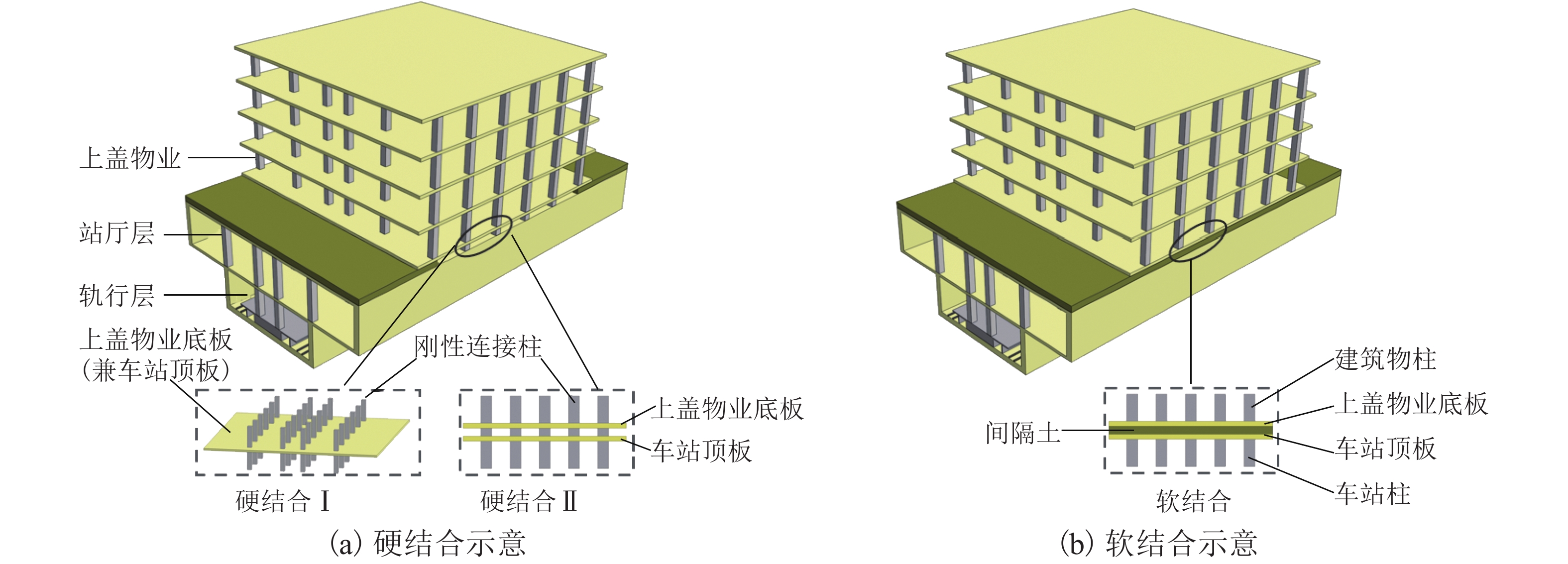
地铁车站与上盖物业的结合型式主要有层间夹土和板、柱等结构直接相连2种型式,在此将其分别归为“软结合”“硬结合”. 其中,“软结合”是上盖物业与地铁车站结构不直接相连,二者之间存在回填或原状夹层,两结构间无刚性连接. “硬结合”是上盖物业与地铁车站间采用刚性连接,具体又分为:1) 硬结合Ⅰ,物业首层作为地铁车站顶板(板共用);2) 硬结合Ⅱ,柱网结合型式(板不共用). 2种结合型式如图1所示.
2. 动力响应分析方法
2.1 列车荷载的确定
列车-轨道振动系统动力学模型分析法是利用解析法或者有限元数值法求解列车荷载,以轮轨不平顺视为车辆振动的激励源,并通过轮轨间赫兹接触关系将其传递到轨道结构中,从而形成整个车轨系统的动力学耦合过程. 车辆-轨道垂向耦合振动的动力模型[21]包括车辆模型、轨道模型和轮轨模型[22]. 车辆模型包括车体、2个转向架以及2组轮对,共计10个自由度.
2.2 数值仿真模型及参数
依托工程为佛山金融高新地铁站,该站为地下2层双柱双跨岛式站台,上盖物业上方有商场及裙楼,车站于下方斜穿上盖建筑,轨道为无砟形式. 采用FLAC3D建立数值仿真模型,鉴于动力计算的难度和耗时,仅考虑受列车振动影响大的上部物业结构. 为减小模型边界效应,确定模型长121.4 m、宽64 m、高72.5 m,地铁车站位于整体模型纵向中心位置. 为精确模拟波的传播,模型网格尺寸应小于输入波形最高频率对应波长的1/8~1/10[23]. 在轨行区、站厅层(A0)及上盖物业(A1~A4)楼板设置25个测点,模型、测点位置及编号见图2.
周边岩土体采用理想弹塑性模型并服从Mohr-Coulomb屈服准则[24],车站结构与上盖物业[25]均为线弹性模型[26]. 动力分析中选用静态边界条件,可有效减小模型边界上的入射波影响[27]. 具体做法是在模型的法向与切向分别设置阻尼器,从而实现吸收入射波的目的.
阻尼器所提供的法向黏性力tn和切向黏性力ts分别为
{tn=−ρCpvn,ts=−ρCsvs, (1) 式中:vn、vs分别为模型边界上法向和切向速度分量,m/s;ρ为介质密度,kg/m3;Cp、Cs分别为P波和S波的波速,m/s.
根据地铁站周边地层勘察资料,车站采用C30混凝土,上盖物业采用C25混凝土,地层、轨道和相关结构的物理力学参数取值如表1和表2所示.
表 1 地层及结构物理力学参数Table 1. Physical and mechanical parameters of strata and structures序号 名称 重度/(KN·m−3) 弹性模量/MPa 泊松比 厚度/m 内摩擦角/(°) 黏聚力/kPa 1 素填土 16.5 18.00 0.33 3.5 5.7 9.4 2 淤泥质粉细砂 17.1 30.00 0.25 6.0 23.0 3 粉质黏土 19.8 105.00 0.32 2.5 22.6 22.2 4 全风化粉砂岩 19.3 225.00 0.29 38 49.3 10.3 5 钢轨 78.5 2.01×10⁵ 0.30 6 地铁车站 24.0 3.00×10⁴ 0.20 7 上盖物业 24.0 2.80×10⁴ 0.20 表 2 轨道部件物理力学参数Table 2. Physical and mechanical parameters of track components钢轨 扣件 道床板 质量/
(kg·m−1)密度/
(kg·m−3)弹性
模量/GPa泊松比 垂向刚度/
(MN·m−1)扣件间距/m 弹性模量/
GPa泊松比 密度/
(kg·m−3)60 7850 205.9 0.30 59.2 0.6 32.5 0.24 2400 2.3 阻尼设置
瑞利阻尼C能够较好减弱系统自振模式的振幅,其矩阵计算如式(2)所示.
C=αM+βK, (2) 式中:α、β分别为与质量、刚度成比例的阻尼常数,M、K分别为质量、刚度矩阵.
阻尼常数可通过最小临界阻尼比和最小中心频率确定. 岩土材料临界阻尼比一般是2%~5%[24],而结构系统的临界阻尼比一般是2%~10%[28]. 模型基本构架大多为矩形块体,相对简单,可采用自振频率作为中心频率. 最终,通过试算得到中心频率为2.1 Hz,土体的质量和刚度阻尼常数分别为0.105、0.024,结构系统的质量和刚度阻尼常数分别为0.042、0.010.
2.4 列车荷载移动化
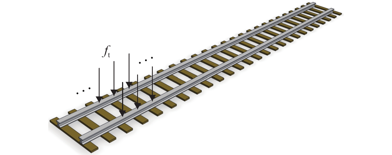
移动荷载与静载对结构的动力响应存在很大区别[29],在列车运行时,经过轨道上不同位置扣件的反力时程曲线在波形上除相位存在差异外,荷载时程曲线及频谱特性基本一致,该相位差的存在是由于列车通过相邻两观测点的时刻不同所导致的. 因此,将前述计算得出的列车荷载作用在钢轨上,施加移动的轮轨作用力. 首先,根据车速和沿列车行进方向单元的长度,计算出列车通过一个单元所需要的时间Δt,再将Δt除以动力时间步长,得到相邻单元所间隔的时间步数,进而实现对不同位置单元施加移动列车荷载. 荷载施加如图3所示,图中,ft为列车振动荷载.
3. 计算方法验证
3.1 现场测试
为验证列车荷载确定方法和构建数值模型的准确性,对依托工程的地铁列车振动进行了现场测试,将数值仿真结果与该地铁站测试数据进行时域和频域的对比分析. 振动信号采集仪器主要有加速度传感器及TST5912动态信号测试分析系统. 测试站点位于该站负1层的站厅层(A0)及上盖物业1~4层(A1~A4),安排专人负责采集列车经过时的振动信号.
3.2 计算模型与验证
该地铁站与上方裙楼及物业的连接方式是以物业首层楼板作为地铁站顶板,故采用2.2节“网格划分”给出的数值模型,仅去除掉软结合夹层,边界条件与材料阻尼参数均与2.2节和2.3节相同,数值仿真模型如图4所示.
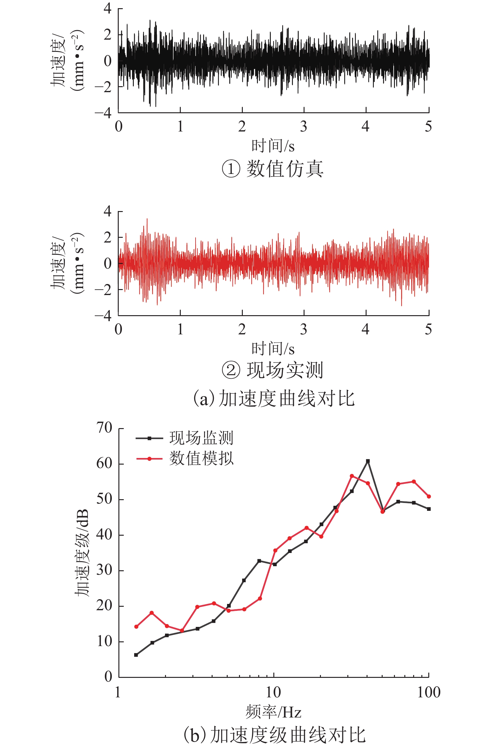
结合列车速度信息,选取现场实测上盖物业1层(A1)加速度的时程数据,与列车速度40 km/h时数值仿真结果进行对比,并将两者加速度数据进行1/3倍频程转换,对现场实测与数值仿真的加速度级进行对比,结果如图5所示.
通过两者的对比分析可以看出:现场实测A1最大振动加速度为3.38 mm/s2,数值仿真计算站厅层最大振动加速度达到3.09 mm/s2,二者的误差为0.29 mm/s2;现场实测和数值仿真的1/3倍频程谱曲线贴合度较高,仅部分频率存在一定差异. 鉴于现场实测不可避免地会受到环境、设备及行人等影响,少量偏差在所难免,但二者数据分布基本一致,可以认为数值仿真方法与计算参数具有良好的可靠性.
4. 仿真结果分析
在数值仿真方法可行、计算参数合理的基础上,建立软结合、硬结合Ⅰ型、硬结合Ⅱ型3种型式下地铁车站上盖物业数值仿真模型,从时域、频域以及动应力出发,研究地铁列车以最大设计速度80 km/h运行时,上盖物业车致振动影响及差异.
4.1 振动加速度时程分析
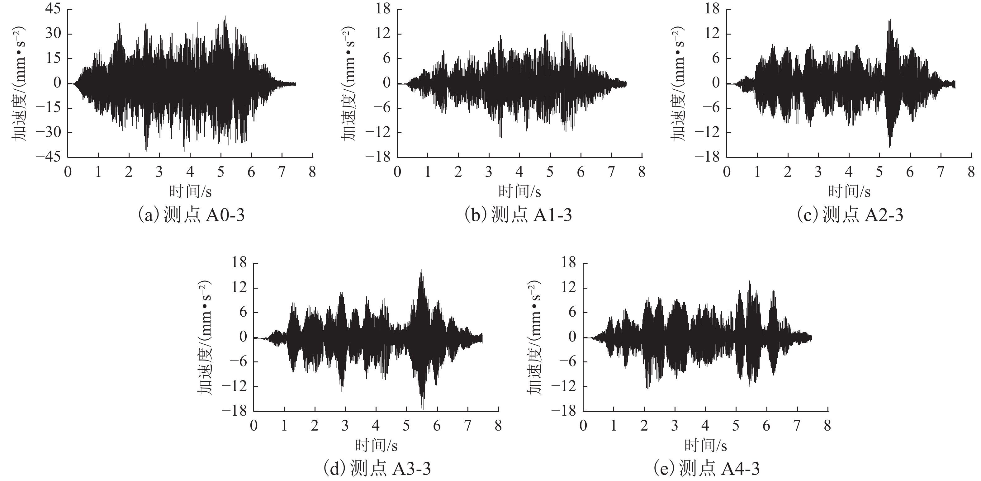
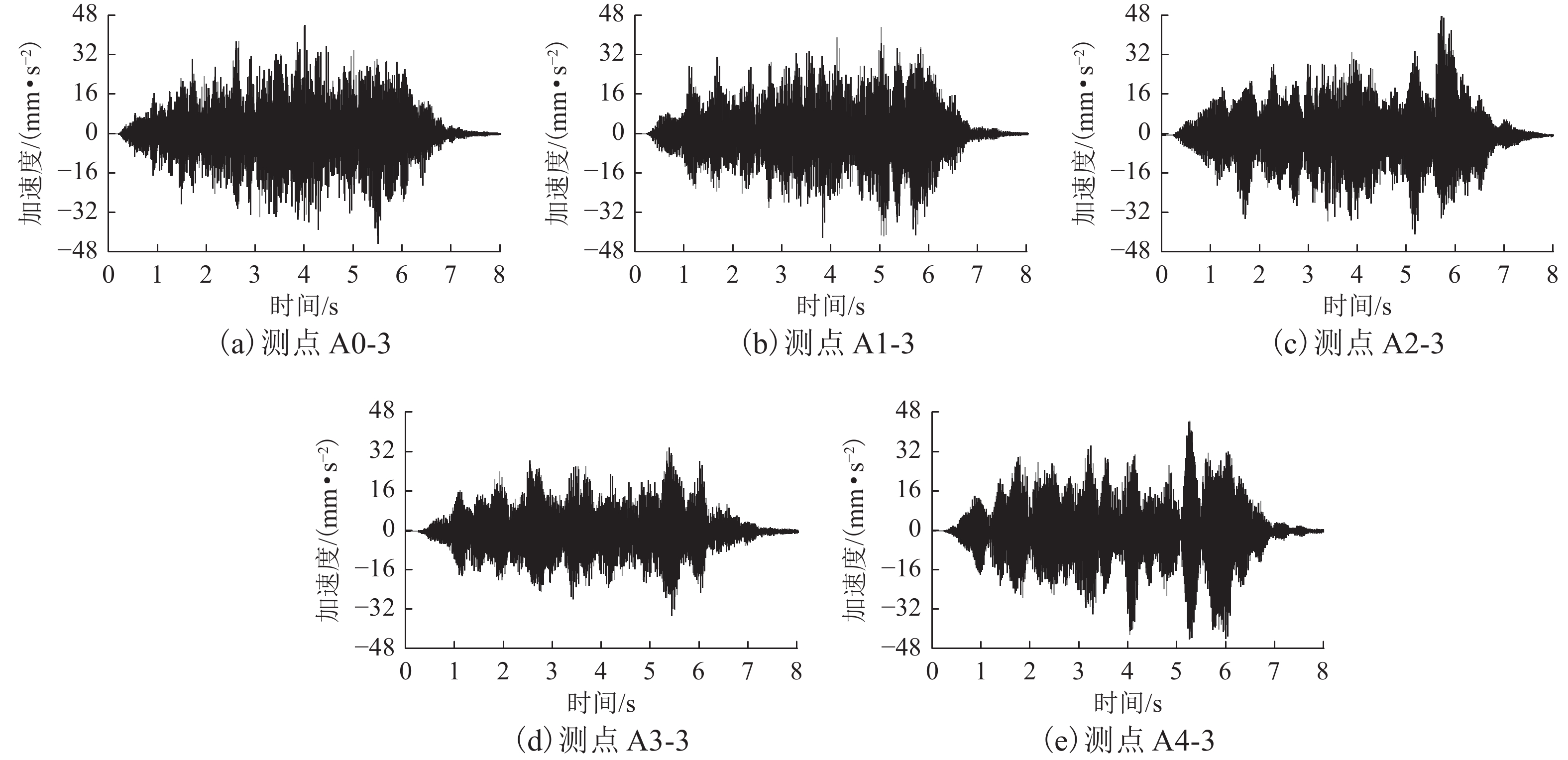
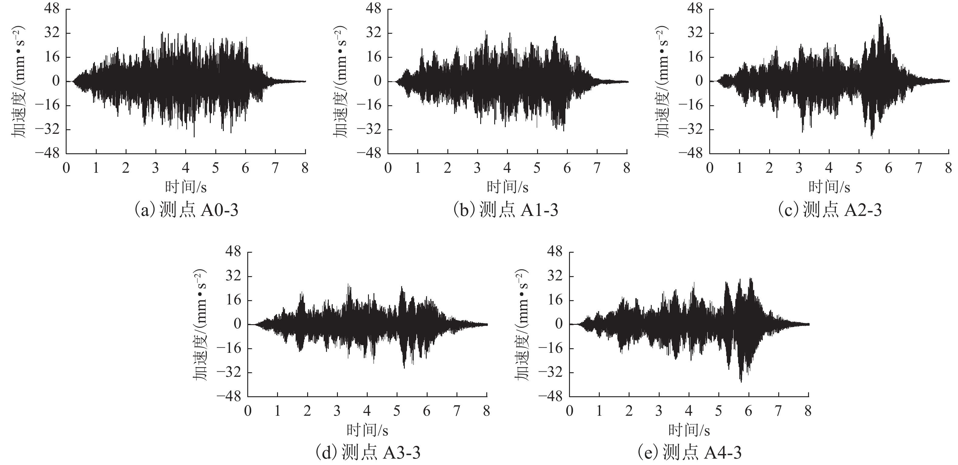
限于篇幅,以断面3的测点振动数据为例进行分析,其振动加速度时程曲线如图6~8所示.
由图6~8可看出:1) 随着列车接近、穿越并驶离建筑物,振动加速度呈现明显的先增大、后减小的规律. 2) 软结合型式下,站厅层A0振动加速度峰值最大,为41.20 mm/s2,上盖物业A1~A4振动加速度峰值分别为12.73、15.61、16.78、13.78 mm/s2,A0到A1振动加速度峰值衰减了69.10%;硬结合Ⅰ型下,A0振动加速度峰值为43.84 mm/s2,A1~A4分别为42.93、50.36、33.56、44.26 mm/s2,A0到A1振动加速度峰值减小了2.08%;硬结合Ⅱ型下,A0振动加速度峰值为32.68 mm/s2,A1~A4分别为33.64、43.47、27.13、30.92 mm/s2,A0到A1振动加速度峰值增大了2.94%. 3) 软结合型式下,上盖物业各楼层振动加速度整体小于硬结合型式,且软结合型式A0到A1加速度衰减较大,而硬结合型式下变化很小,说明软结合型式的间隔土对列车振动有削减作用. 4) 硬结合Ⅰ型各楼层振动加速度略大于硬结合Ⅱ型,原因是硬结合Ⅱ型的车站-上盖物业刚度更大.
4.2 频域下动力响应分析
依据断面3各测点振动加速度的计算结果,采用快速傅里叶变换将时域结果转换到频域,通过MATLAB得到1/3倍频程各中心频率的振动加速度级. 3种结合型式下各测点加速度级峰值曲线如图9所示.
3种结合型式下各测点加速度级峰值曲线如图9所示. 以断面3为例,软结合型式下地铁车站上盖物业加速度级最大值位于A0,为79.5 dB,A1的值为68.2 dB,较A0减小了11.3 dB,说明地铁车站与上盖物业结构间的间隔土对列车振动有较大的衰减作用;除A1外,各楼层加速度级最大值均位于断面3,加速度级从断面3往两侧逐渐减小,说明对于同一楼层,随着距振源距离的增大振动减小. 硬结合Ⅰ型与硬结合Ⅱ型地铁车站上盖物业加速度级水平与竖向分布规律基本相同;对于上盖物业同一楼层,均是断面3加速度级最大,往两侧逐渐减小,说明随着距振源距离的增大振动减小;地铁车站上盖物业加速度级最大值均位于A4,硬结合Ⅰ型最大值为83.4 dB,硬结合Ⅱ型最大值为79.4 dB,均较A0有所增大. 硬结合型式下,上盖物业加速度级整体上较软结合大,并且硬结合Ⅰ型大于硬结合Ⅱ型. 因此,在条件允许的情况下,地铁车站上盖物业优先选择软结合型式,其次选择硬结合Ⅱ型.
图10为3种结合型A1各断面振动加速度频谱,图11为3种结合型断面3各楼层的振动加速度频谱. 由图10、11可以看出:3种结合型,上盖物业车致振动响应频率的分布规律基本一致,上盖物业车致振动频率主要集中在40~90 Hz;对于同一断面,各楼层振动响应频率范围及振幅变化不大.
4.3 动应力分析
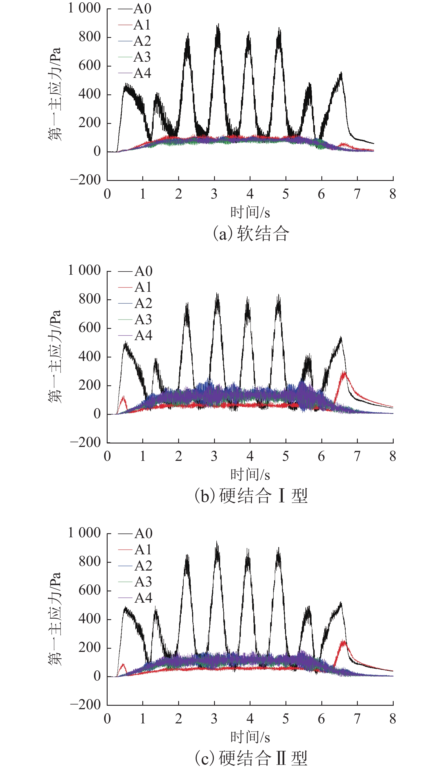
3种结合型式下,断面3各楼层测点第一主应力时程曲线如图12所示,各主应力峰值如表3所示.
从图12、表3可以看出:软结合、硬结合Ⅰ型、硬结合Ⅱ型的第一主应力最大值均位于A0,分别为899.33、849.54、952.66 Pa,A1~A4第一主应力最大值远小于A0最大值;软结合型式第一主应力从A0到A1衰减了85.81%,衰减较大,主要是车站与上盖物业间隔土削弱了列车振动,进而减小了列车荷载作用下上盖物业的附加应力;硬结合Ⅰ、Ⅱ型第一主应力从A0到A1分别衰减了63.46%、72.27%,比软结合型衰减慢,主要是因为车站与上盖物业刚性连接,列车振动可以通过连接柱向上传播.
表 3 不同结合型断面3各楼层的第一主应力峰值Table 3. First principal stress peak value of the third section of each floor under different combination types楼层 软结合 硬结合Ⅰ 硬结合Ⅱ A0 899.33 849.54 952.66 A1 127.60 310.39 264.16 A2 113.31 264.24 180.05 A3 109.44 200.76 172.24 A4 120.30 233.98 192.38 衰减率/% 85.81 63.46 72.27 注:衰减率指A0到A1的第一主应力衰减率. 5. 结 论
采用现场测试和数值仿真手段对提出的地铁车站-上盖物业间的软结合、硬结合Ⅰ型和硬结合Ⅱ型3种型式的车致振动响应差异进行了研究,得到如下结论:
1) 依托现场实测数据验证了数值仿真方法与计算参数的可靠性,两者的A1最大振动加速度差值仅为0.29 mm/s2,且1/3倍频程谱曲线分布也基本一致.
2) 从A0到A1,振动加速度峰值软结合型减小了69.10%,硬结合Ⅰ型减小了2.08%,硬结合Ⅱ型增大了2.94%;硬结合型式下上盖物业振动加速度较软结合型式大.
3) 3种结合型式下,上盖物业振动的频率主要在40~90 Hz,且对于上盖物业同一楼层,车致振动随着距振源距离的增大而逐渐减小;软结合型A1加速度级最大值为68.2 dB,较A0减小11.3 dB;硬结合型上盖物业加速度级整体较软结合大,硬结合Ⅰ型上盖物业加速度级最大值为83.4 dB,硬结合Ⅱ型为79.4 dB.
4) 地铁列车振动造成上盖物业附加第一主应力很小,且在向上传播过程中衰减很快;从A0到A1,软结合型、硬结合Ⅰ型、硬结合Ⅱ型分别衰减85.81%、63.46%、72.27%,间隔土对附加应力有明显衰减作用.
5) 在相同列车荷载作用下,列车振动对软结合型地铁车站上盖物业影响最小,其次为硬结合Ⅱ型,最后为硬结合Ⅰ型;实际工程中,如果现场条件允许,建议优选软结合型式.
-
表 1 不同主干网络分类精度对比
Table 1. Comparison of classification accuracy for different backbone networks
% 网络模型 训练集 测试集 精度 F1 分数 精度 F1 分数 ResNet18 83.5 82.0 82.3 81.7 ResNet34 85.6 84.9 84.8 82.3 ResNet50 90.8 87.6 81.2 76.3 VGG-16 80.9 79.8 80.6 79.6 VGG-19 82.5 81.6 80.0 79.5 表 2 分块尺度对比实验
Table 2. Comparison experiments of partition scales
% 块尺度
大小/块训练集 训练集 精度 F1 分数 精度 F1 分数 16 83.1 82.9 82.8 82.3 8 84.2 83.9 83.2 82.5 4 89.6 89.3 88.3 87.7 2 86.1 85.7 85.3 85.1 表 3 图像打乱对比实验
Table 3. Comparison experiments of shuffled images
% 打乱方法 训练集 测试集 精度 F1 分数 精度 F1 分数 未打乱 85.6 84.9 84.8 82.3 随机打乱 88.2 87.9 87.2 86.5 分区打乱 89.6 89.3 88.3 87.7 表 4 重校准损失函数对分类精度的影响
Table 4. Influence of recalibration loss function on classification accuracy
% 损失函数 训练集 测试集 精度 F1 分数 精度 F1 分数 Log-Cosh 91.7 91.6 90.3 90.8 MAE 88.3 88.5 86.1 86.7 SMAE 89.3 89.1 90.6 90.2 MSE 93.8 92.5 91.3 91.7 表 5 重校准损失函数和分类损失函数的权重对分类精度的影响
Table 5. Influence of weights of recalibration loss function and classification loss function on classification accuracy
% 损失函数权重 训练集 测试集 Ls Lcla 精度 F1 分数 精度 F1 分数 1.00 1.00 93.8 92.5 91.3 91.7 0.25 0.75 90.3 89.7 89.3 88.3 0.50 0.50 91.5 90.8 90.6 90.2 0.75 0.25 92.3 91.2 90.8 90.5 表 6 不同融合方法对分类精度的影响
Table 6. Influence of different fusion methods on classification accuracy
% 融合方法 训练集 测试集 精度 F1 分数 精度 F1 分数 Max 93.4 92.9 92.7 92.2 Sum 92.6 92.0 92.3 91.7 Conv 94.9 94.5 94.3 93.6 表 7 不同打乱方法和重校准损失函数下的消融实验
Table 7. Ablation experiments with different shuffle methods and recalibration loss functions
% 打乱方法 损失函数 训练集 测试集 精度 F1 分数 精度 F1 分数 未打乱 Log-Cosh 86.2 85.3 84.8 83.3 MAE 85.2 84.9 83.7 83.1 SMAE 85.6 84.7 84.2 83.9 MSE 86.8 86.2 85.9 85.7 随机打乱 Log-Cosh 87.2 86.9 87.2 86.3 MAE 86.2 86.9 86.2 86.3 SMAE 86.7 86.3 85.8 85.6 MSE 87.9 87.3 87.1 86.5 分区打乱 Log-Cosh 91.7 91.6 90.3 90.8 MAE 88.3 88.5 86.1 86.7 SMAE 89.3 89.1 90.6 90.2 MSE 93.8 92.5 91.3 91.7 表 8 BUSI数据集实验结果
Table 8. Experimental results of BUSI dataset
% 融合方法 训练集 测试集 精度 F1 分数 精度 F1 分数 Max 93.4 92.9 92.7 92.2 Sum 92.6 92.0 92.3 91.7 Conv 94.9 94.5 94.3 93.6 表 9 本文模型与主流方法性能比较
Table 9. Performance comparison between proposed method and popular methods
-
[1] SUNG H, FERLAY J, SIEGEL R L, et al. Global cancer statistics 2020: GLOBOCAN estimates of incidence and mortality worldwide for 36 cancers in 185 countries[J]. CA: a Cancer Journal for Clinicians, 2021, 71(3): 209-249. doi: 10.3322/caac.21660 [2] HOOLEY R J, SCOUTT L M, PHILPOTTS L E. Breast ultrasonography: state of the art[J]. Radiology, 2013, 268(3): 642-659. doi: 10.1148/radiol.13121606 [3] 龚勋,杨菲,杜章锦,等. 甲状腺、乳腺超声影像自动分析技术综述[J]. 软件学报,2020,31(7): 2245-2282.GONG Xun, YANG Fei, DU Zhangjin, et al. Survey of automatic ultrasonographic analysis for thyroid and breast[J]. Journal of Software, 2020, 31(7): 2245-2282. [4] LO C M, CHANG Y C, YANG Y W, et al. Quantitative breast mass classification based on the integration of B-mode features and strain features in elastography[J]. Computers in Biology and Medicine, 2015, 64: 91-100. doi: 10.1016/j.compbiomed.2015.06.013 [5] FLORES W G, DE ALBUQUERQUE PEREIRA W C, INFANTOSI A F C. Improving classification performance of breast lesions on ultrasonography[J]. Pattern Recognition, 2015, 48(4): 1125-1136. doi: 10.1016/j.patcog.2014.06.006 [6] 任丽,刘洋洋,童莹,等. 乳腺肿瘤超声图像的多特征提取及分类研究[J]. 中国医疗器械杂志,2020,44(4): 294-301.REN Li, LIU Yangyang, TONG Ying, et al. Multi-feature extraction and classification of breast tumor in ultrasound image[J]. Chinese Journal of Medical Instrumentation, 2020, 44(4): 294-301. [7] SPANHOL F A, OLIVEIRA L S, PETITJEAN C, et al. Breast cancer histopathological image classification using convolutional neural networks[C]//2016 International Joint Conference on Neural Networks (IJCNN). Vancouver: IEEE, 2016: 2560-2567. [8] WEI B Z, HAN Z Y, HE X Y, et al. Deep learning model based breast cancer histopathological image classification[C]//2017 IEEE 2nd International Conference on Cloud Computing and Big Data Analysis (ICCCBDA). Chengdu: IEEE, 2017: 348-353. [9] XIE J, SONG X S, ZHANG W, et al. A novel approach with dual-sampling convolutional neural network for ultrasound image classification of breast tumors[J]. Physics in Medicine and Biology, 2020, 65(24): 245001.1-245001.15. [10] CAO Z T, DUAN L X, YANG G W, et al. An experimental study on breast lesion detection and classification from ultrasound images using deep learning architectures[J]. BMC Medical Imaging, 2019, 19(1): 51.1-51.9. [11] 孔小函,檀韬,包凌云,等. 基于卷积神经网络和多信息融合的三维乳腺超声分类方法[J]. 中国生物医学工程学报,2018,37(4): 414-422.KONG Xiaohan, TAN Tao, BAO Lingyun, et al. Classification of breast mass in 3D ultrasound images with annotations based on convolutional neural networks[J]. Chinese Journal of Biomedical Engineering, 2018, 37(4): 414-422. [12] 迟剑宁,于晓升,张艺菲. 融合深度网络和浅层纹理特征的甲状腺结节癌变超声图像诊断[J]. 中国图象图形学报,2018,23(10): 1582-1593.CHI Jianning, YU Xiaosheng, ZHANG Yifei. Thyroid nodule malignantrisk detection in ultrasound image by fusing deep and texture features[J]. Journal of Image and Graphics, 2018, 23(10): 1582-1593. [13] 杜章锦,龚勋,罗俊,等. 乳腺超声图像中易混淆困难样本的分类方法[J]. 中国图象图形学报,2020,25(7): 1490-1500.DU Zhangjin, GONG Xun, LUO Jun, et al. Classification method for samples that are easy to be confused in breast ultrasound images[J]. Journal of Image and Graphics, 2020, 25(7): 1490-1500. [14] DU R Y, CHANG D L, BHUNIA A K, et al. Fine-grained visual classification via progressive multi-granularity training of jigsaw patches[C]//European Conference on Computer Vision. Cham: Springer, 2020: 153-168. [15] 杨丽娜. 乳腺癌超声图像报告中的BI-RADS与术后病理结果之间的相关性[J]. 世界复合医学,2018,4(6): 51-53.YANG Li’na. Correlation between BI-RADS and postoperative pathological findings in breast cancer ultrasound image reports[J]. World Journal of Complex Medicine, 2018, 4(6): 51-53. [16] LAMPLE G, OTT M, CONNEAU A, et al. Phrase-based & neural unsupervised machine translation[C]//Proceedings of the 2018 Conference on Empirical Methods in Natural Language Processing. Brussels: Association for Computational Linguistics, 2018: 5039-5049. [17] CHEN Y, BAI Y L, ZHANG W, et al. Destruction and construction learning for fine-grained image recognition[C]//2019 IEEE/CVF Conference on Computer Vision and Pattern Recognition (CVPR). Long Beach: IEEE, 2019: 5152-5161. [18] AL-DHABYANI W, GOMAA M, KHALED H, et al. Dataset of breast ultrasound images[J]. Data in Brief, 2020, 28: 104863.1-104863.5. [19] SIMONYAN K, ZISSERMAN A. Very deep convolutional networks for large-scale image recognition[EB/OL]. (2014-09-14)[2021-09-02]. https://ar5iv.labs.arxiv.org/html/1409.1556. [20] HE K M, ZHANG X Y, REN S Q, et al. Deep residual learning for image recognition[C]//2016 IEEE Conference on Computer Vision and Pattern Recognition (CVPR). Las Vegas: IEEE, 2016: 770-778. [21] WEI M W, DU Y Z, WU X M, et al. A benign and malignant breast tumor classification method via efficiently combining texture and morphological features on ultrasound images[J]. Computational and Mathematical Methods in Medicine, 2020, 2020: 5894010.1-5894010.12. [22] ABED MOHAMMED M, AL-KHATEEB B, RASHID A N, et al. Neural network and multi-fractal dimension features for breast cancer classification from ultrasound images[J]. Computers & Electrical Engineering, 2018, 70: 871-882. [23] SHIN S Y, LEE S, YUN I D, et al. Joint weakly and semi-supervised deep learning for localization and classification of masses in breast ultrasound images[J]. IEEE Transactions on Medical Imaging, 2019, 38(3): 762-774. doi: 10.1109/TMI.2018.2872031 -


 下载:
下载:


 下载:
下载:

